Thanks for the response. I did check osimis, it was easy to install.
However I am not able to view any output. I mean the series is opening successfully in osimis but I don’t see any graphical data whereas I see Patient Id, Study Id etc.
I did explore with different menu options available on screen but couldn’t get any output.
Any idea on what can be the issue? If image isn’t in proper format, I shouldn’t even open. Am I missing something?
Thanks
Selva
In general, it is difficult to view images modified from original
DICOM files in general DICOM viewer.
However, there are some solutions for it.
For example, you should export them in RAW format and import them by
your DICOM viewer in the DICOM format giving proper DICOM information
like Patient ID, photometricInterpretation, modality...etc.
In another way, you should customize some DICOM tags associated with
multi frame format like (0028, 0009)... and modify your DICOM viewer
so that it can read the DICOM tags.
I have done such a kind of work in order to segment tissues and
analyze it using FEM from medical images in DICOM.
Actually we are trying to use a platform that will automate the full cycle. I mean from image acquisition from Orthanc to image analysis in 3rd party platform and image results back to Orthanc. So, we would like to view the segmented image in Orthanc.
Okay, but where is it stored or I can see the output images in Orthanc?
As I am not able to view in Orthanc, if I would like to download it and view it in my local desktop, how can I do it?
Hello… I am sorry but it seems I was mistaken (I am rather new to Dicom:) ). The Dicom segmentation image objects, according to an Osimis developer, aren’t supported, neither in the Orthanc viewer nor in the Osimis plugin.
If this is an important part of your application, maybe you should consider arranging for a custom development with an Orthanc commercial partner.
HorliX, my customized version of Horos/OsiriX, can read it as follows.
Maybe OsiriX MD can do so although I don’t have it because it is somewhat expensive for me.
Of course, I respect @rossetantonie and contributors of OsiriX.
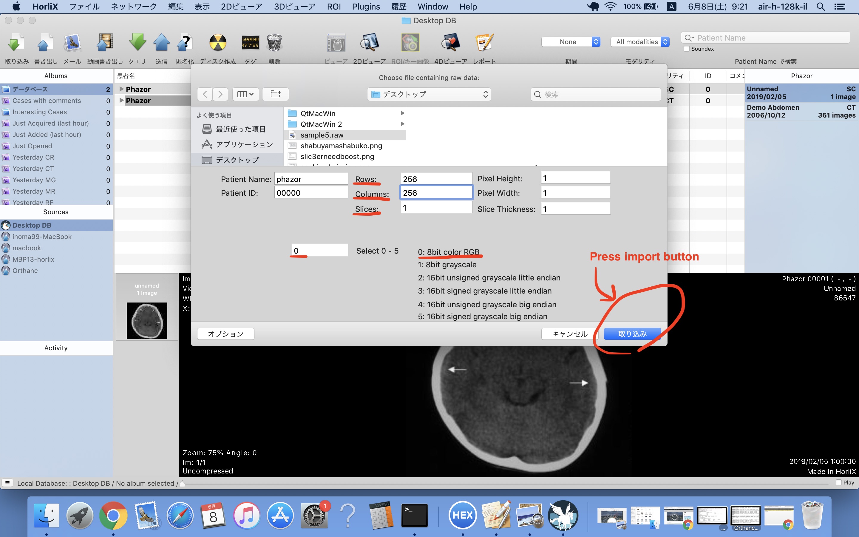
First of all, you select ‘Import Raw Data2…’ menu item like Fig1.jpg .
Dialog Box come up. You specify Rows, Columns, Slices, photometricInterpretation… like Fig2.jpg .
In this case sample file has a single image of 256x256 8bit RGB.
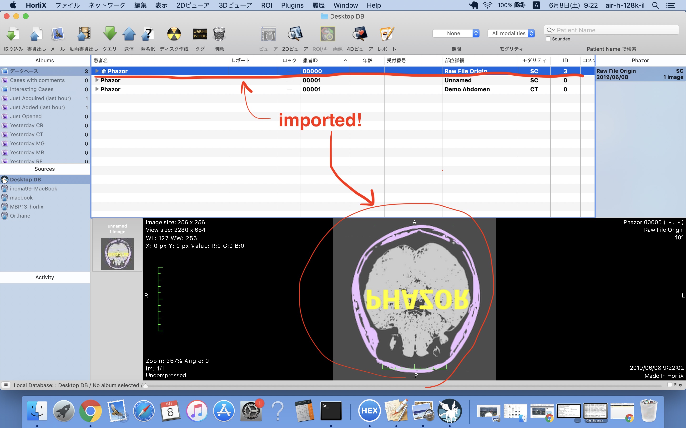
Then HorliX import it in the format you specified like Fig3.jpg .
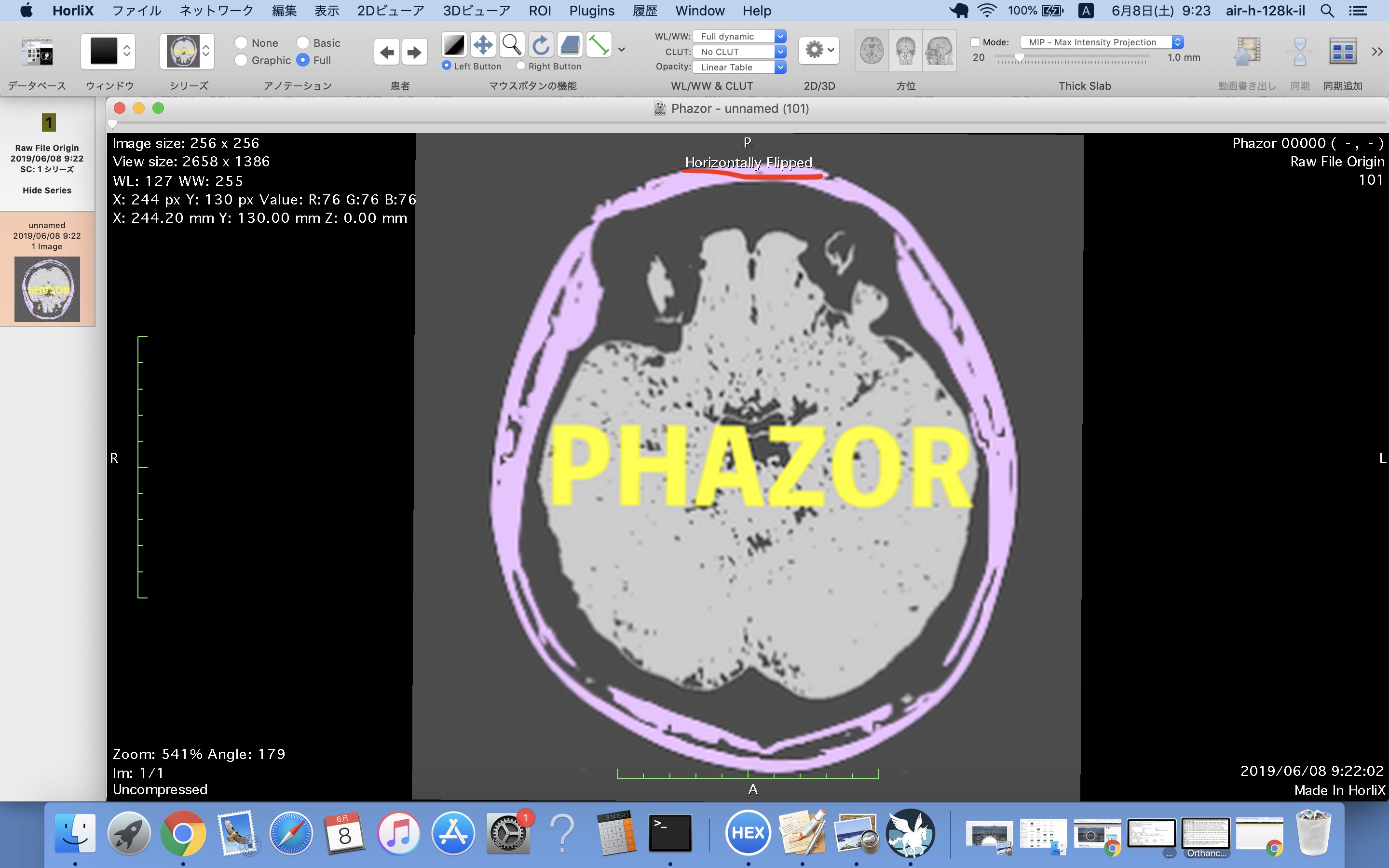
Oh, the image is flipped
Double click the image and 2D Viewer comes up: See Fig4.jpg .
You should adjust the image using rotation and flipping functions as you like.
Hiroaki: Orthanc is about DICOM, not about RAW files that are fully vendor-specific. First convert the RAW files to DICOM before using them in Orthanc.
Selva: Please have a look at the following FAQ entry in the Orthanc Book: